Zebrabow

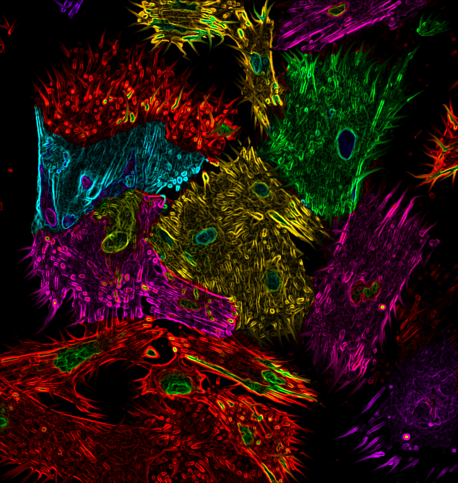
Zebrabow embryos express random combinations of red, green, and blue fluorescent proteins, revealing a spectrum of unique hues. These same hues can be used to ‘barcode’ individual stem cells to track their birth and contribution to tissues as the embryo grows–all cells of the same hue were produced from one stem cell’s divisions. The Zebrabow system allows for long-term tissue lineage analysis, because the fluorescent proteins will … Continue reading Zebrabow

A Sight For Sore Eyes

  Have you ever experienced itchy, watery eyes as a result of seasonal allergies? The objects in this picture are to blame. This microscope image shows conjunctival tissue from an eye in the presence of a single mast cell. Mast cells are important regulators of the immune system. Upon invasion of the eye with foreign objects, such as allergens, the mast cell is recruited to … Continue reading A Sight For Sore Eyes

Holding the Fort Down

And how! Depicted here are intricate collagen fibers, the structural glue and the most abundant protein in the mammalian body. Collagen proteins occupy the extracellular space in connective tissues, and form fibers that provide the physical scaffold for holding and strengthening several tissues in the body including bones, ligaments, tendons, skin, and the cornea. With several different classes of this protein defined to date, type … Continue reading Holding the Fort Down

Branched synaptic arbor in fillet

The Drosophila neuromuscular junction is a beautiful and yet powerful model synapse for in vivo studies of development, physiology, cell biology and plasticity. This illustration by Saskia Van Vactor shows a field of ventral longitudinal body wall muscles in red, as if stained with phalloidin to highlight filamentous actin, the main protein building block of muscle. Against this background, several branches of intersegmental nerve extend … Continue reading Branched synaptic arbor in fillet

The Brain is a Series of Tubes

The brain is made of up billions of neurons and even more connections between neurons. We can get an idea of how neurons are connected across the human brain using a type of brain scan called diffusion-weighted magnetic resonance imaging (dMRI). How does dMRI work? It actually measures the direction that water moves throughout the brain. Sometimes, water can move in all directions equally easily – like … Continue reading The Brain is a Series of Tubes

Retinal Corona

Image contributed by Yu Wang, a third year graduate student at Harvard Medical School. Glial cells support the proper function of neuronal cell types and are also responsible for cleaning up cell debris such as leftover neurotransmitter molecules (what neurons use to communicate with each other). Certain types of glia, called microglia, function as the first line of defense in immune-privileged places such as the … Continue reading Retinal Corona

Cryolysed

This is a layer of epithelial cells that was subjected to sever freeze-thaw cycles before being imaged at 400x magnification, which caused the membranes to rupture and form these arrays of crystal-like structures. When frozen, the water inside the cells expands, breaking through the cell membrane. With the membrane integrity compromised, when the cells are thawed the water inside leaks out, the fragments of membrane form aggregates, … Continue reading Cryolysed

Tubular

These images show cross sections of the different compartments of the developing gut in a chick embryo: esophagus (top), small intestine (middle), and large intestine (bottom), at days 6, 8, 10, and 14 (left to right). As the chick develops, the shape of the gut changes from a simple tube to a more complex form with specific surface folding patterns. The way the surface is … Continue reading Tubular